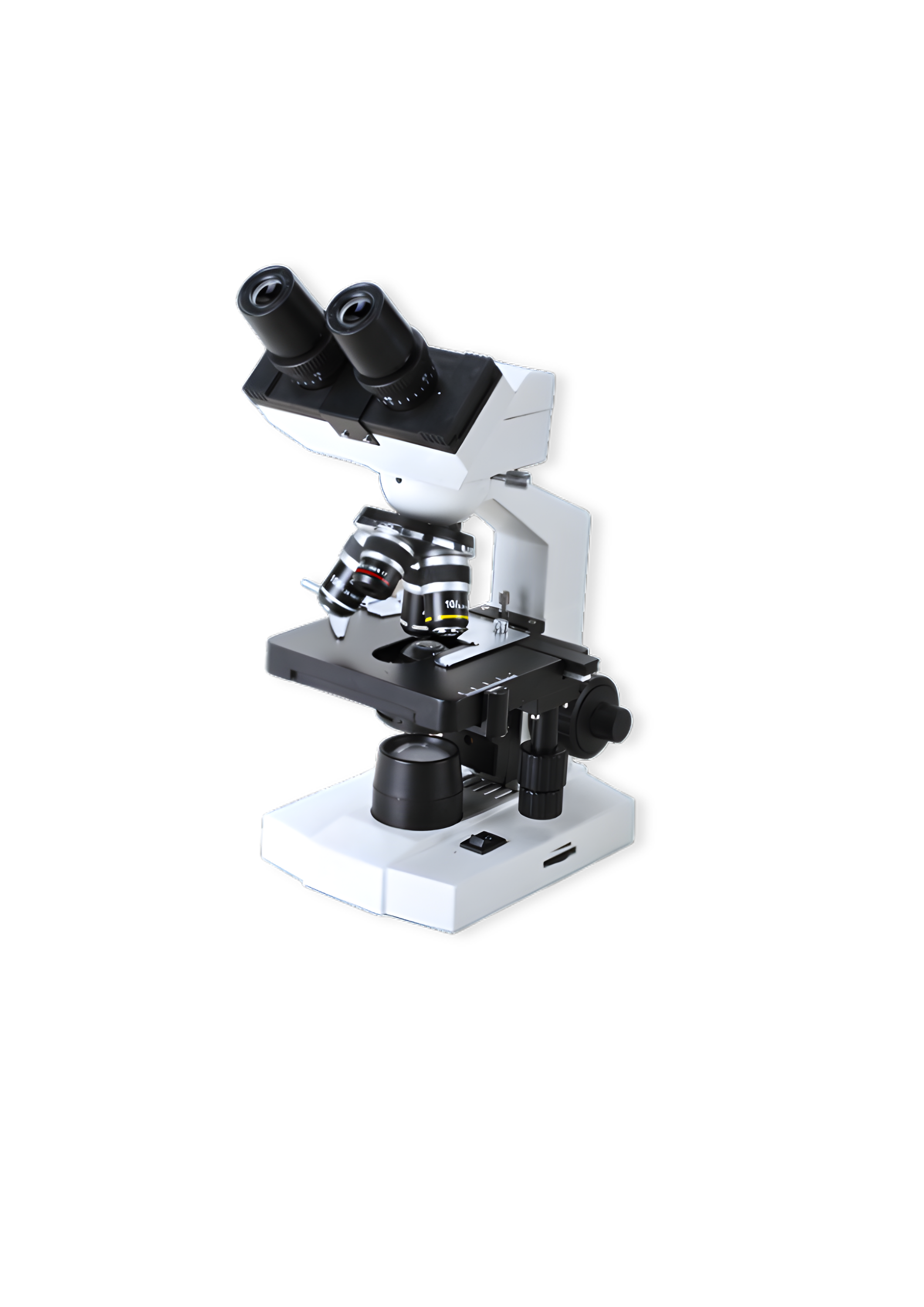
Microscopes - Pathological and Research
Microscopes - Pathological and Research is designed to provide precision, durability, and ease of use for laboratories, educational institutions, and medical facilities. With advanced optics and ergonomic features, this microscope ensures high-resolution imaging and prolonged operational efficiency. It is equipped with anti-reflection and antifungal coatings to enhance clarity and longevity.
Precision Optical Performance
High-quality optical system with plan achromatic objectives ensures sharp, high-contrast, and distortion-free imaging
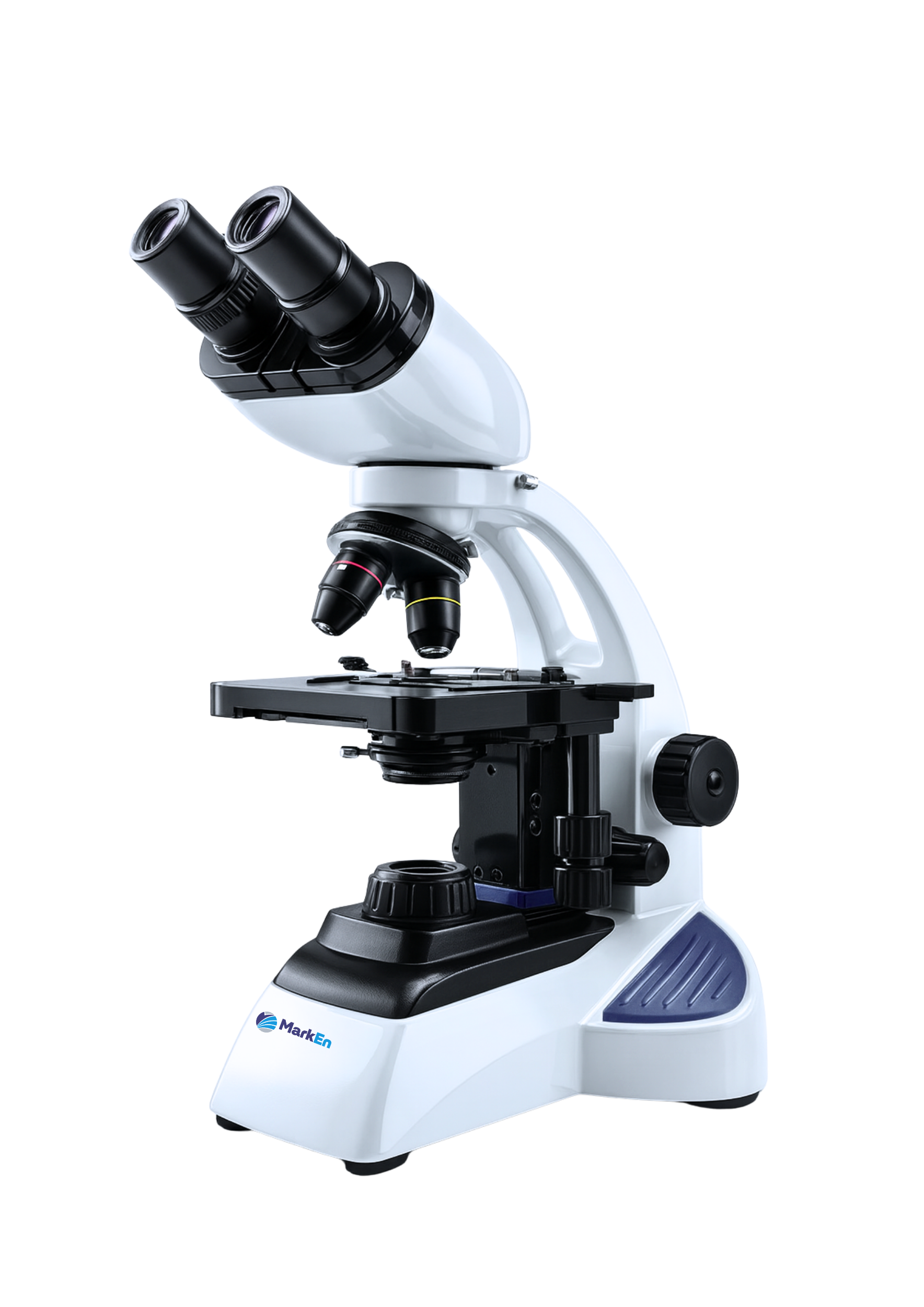
Ergonomic Binocular Viewing Design
30° inclined, 360° rotatable binocular head with adjustable interpupillary distance provides comfortable viewing.
Advanced LED Illumination System
Integrated high-intensity LED illumination delivers bright, uniform lighting with low heat generation.
Smooth & Accurate Focusing Mechanism
Coaxial coarse and fine focusing controls offer smooth, precise specimen positioning with excellent stability and repeatability.
Heavy-Duty Mechanical Stage Assembly
Precision-engineered stage with anti-collision protection enables secure slide handling.
Robust & Long-Life Construction
Solid, vibration-resistant build designed for continuous laboratory operation with long-term reliability.
Make | MarkEn |
Microscope Type | Pathological binocular, non-hinged type with in-built illumination and light intensity regulator |
Observation Tube | Binocular, 30° inclined, 360° rotatable; anti-reflection optical coating. |
Body Construction | Die-cast aluminium body with hexagonal base; precision ball-bearing and wire guide movement |
Optical System | Parallel/Greenough or Infinity Corrected System |
Nosepiece | Revolving 5-position coded nosepiece |
Objective Lens | Plan Achromatic Antifungal Lens. |
Objectives | Plan-Achromat: 5x/0.12, 10x/0.25, 20x/0.45, 40x/0.65 (Spring), 100x/1.25 Oil |
Antifungal & Anti-reflection Coatings | Certified by spectrophotometry report/self-declaration on manufacturer letterhead. |
Stage | 75 × 30 mm mechanical stage with anti-collision mechanism and torque-set fine focusing |
Anti-collision nosepiece | Revolving 5-position encoded nosepiece with anti-collision mechanism |
Stage Clips | High-tension clips for firmly holding microscope slides in place. |
Magnification Range | 6.3x – 55x or higher; coded zoom; distortion-free (≥9:1 ratio) |
Specimen Holder | Spring-loaded, one-hand operation |
Condenser | 5-Position turret condenser (Brightfield, Phase, Darkfield) |
Camera System | Optional/integrated 12 MP CMOS camera (4032 × 3044 px) with 4K/60 fps output through HDMI, LAN, USB 3.0, and Type-C; includes 1/2.3" CMOS sensor (1.85 × 1.85 µm pixel size) with auto image settings, measurement tools, movie recording, and step-less scaling; operates without any adapter |
Illumination | ≥10 W LED with uniform, transmitted/reflected lighting; ≥60,000-hour life; integrated power unit; auto switch-off after 15 minutes; consistent brightness at all magnifications without manual adjustment, Light Manager for TL & RL, compatible with LED and HAL, HAL illumination compatibility included |
Software | Grain size analysis, 2D CAD overlay, 2.5D topography; metadata management; tiles panorama; EDF, management, visualization and printing of images and metadata |
Image Capture & Storage | Snap buttons on both sides of stand for instant image capture; integrated memory card slot for saving images and videos |
Computer (Optional) | i5/i7, 16GB RAM, 512GB SSD, 1GB GPU, Win-10, direct display on HDMI monitor without PC |
Wireless control | System supports wireless control via tablet / PC / laptop |
Optional Upgrades | Camera attachment, Phase Contrast, Fluorescence Head |
Power Supply | 100–240 V AC, 50–60 Hz |